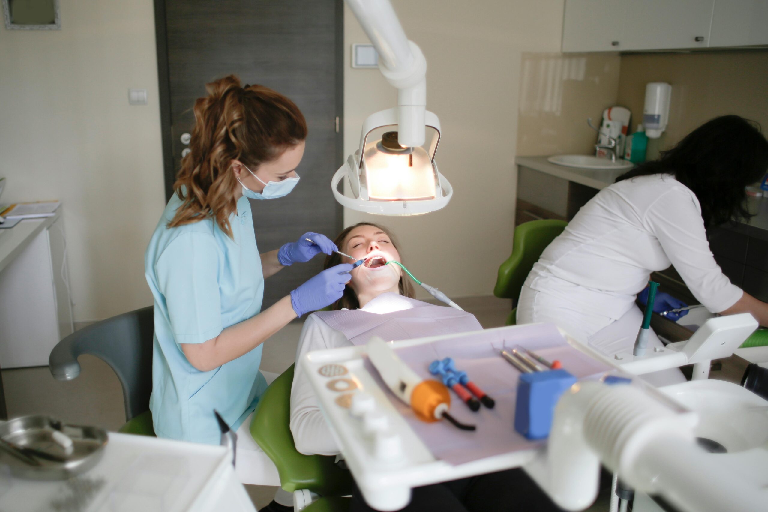
Service
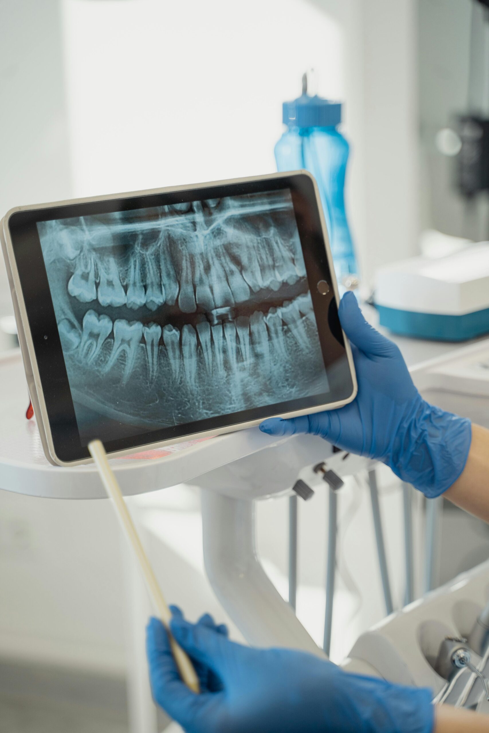
DIGITAL IMAGING (X-RAYS)
Diagnose with confidence using our low-radiation digital X-rays and 3D imaging in Singapore. Immediate results for precise treatment planning.
Book Online
High-Resolution Digital Imaging & X-Rays
Digital imaging has revolutionized modern dentistry, providing immediate, high-definition views of teeth, bone structures, and soft tissues – often with up to 80% less radiation than traditional film. Our clinic employs state-of-the-art sensors and cone-beam computed tomography (CBCT) to diagnose issues invisible to the naked eye, plan implant placements, assess jaw pathology, and monitor treatment outcomes with unprecedented accuracy.
Why Digital Imaging Matters
- Early Detection: Identify interproximal decay, root fractures, and bone loss before symptoms arise.
- Enhanced Treatment Planning: 3D CBCT data lets us virtually place implants, assess sinus proximity, and evaluate nerve location.
- Patient Education: Instantly view and annotate images on-screen, guiding you through diagnoses and treatment options.
- Lower Radiation: Modern digital sensors require significantly less X-ray exposure, enhancing patient safety.
Imaging Modalities & Indications
1. Intraoral Bitewing X-Rays
- Use: Detect decay between back teeth (premolars/molars).
- Benefit: Small sensors capture precise interproximal detail.
2. Periapical X-Rays
- Use: View entire tooth from crown to root tip; assess root health and periapical pathology.
- Benefit: Essential for diagnosing abscesses, root resorption, and periodontal bone levels.
3. Panoramic Radiograph
- Use: Full-arch overview including all teeth, jaws, sinuses, and TMJ.
- Benefit: Ideal for wisdom tooth evaluation, jaw fractures, and broad pathology screening.
4. Cone-Beam Computed Tomography (CBCT)
- Use: 3D imaging for implant planning, impacted teeth, airway analysis, and complex endodontic cases.
- Benefit: Volumetric data with fine isotropic voxels (0.08-0.2 mm resolution), allowing multiplanar reconstruction.
5. Cephalometric Radiograph (Orthodontic Only)
- Use: Lateral skull view to analyze jaw relationships and growth patterns.
- Benefit: Critical for orthodontic diagnosis and surgical planning.
Benefits & Patient Comfort
-
- Comfort: No uncomfortable film; sensors are thinner and more flexible.
-
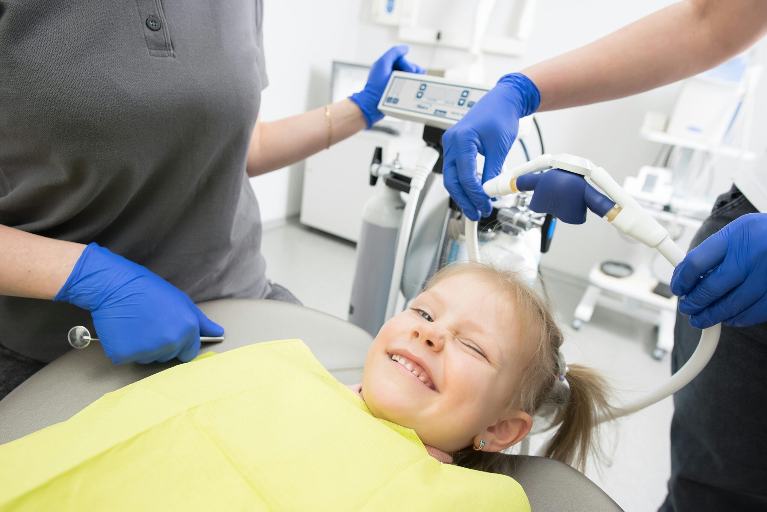
- Safety: Up to 80% less radiation than film, with adjustable exposure settings for children.
-
- Efficiency: Immediate digital files streamline referrals and lab communications.
- Record Keeping: Digital archives allow longitudinal comparisons to track disease progression or healing.